A Brief History of Advanced Normalization Tools (ANTs)
Brian B. Avants (PENN) and
Nicholas J. Tustison (UVA)
This talk is online at http://stnava.github.io/ANTsTalk/ with colored links meant to be clicked for more information
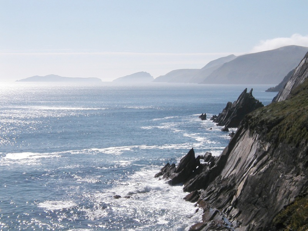
Image mapping & perception: 1878
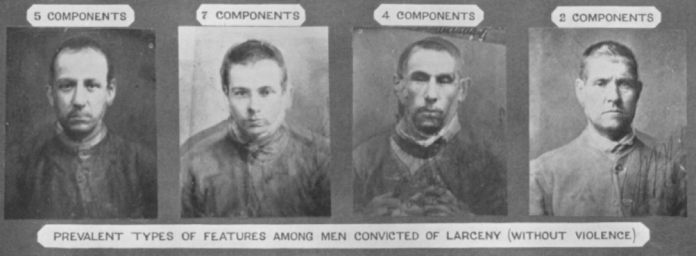
Francis Galton: Can we see criminality in the face?
(maybe he should have used ANTs?)
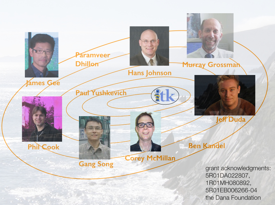
Founding developers
Long-term collaborators
\(+\) neurodebian, slicer, brainsfit, nipype, itk and more …
ANTs Lineage
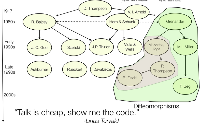
References: Horn and Schunck (1981), Gee, Reivich, and Bajcsy (1993), Grenander (1993), Thompson et al. (2001), Miller, Trouve, and Younes (2002), Shen and Davatzikos (2002), Arnold (2014), Thirion (1998), Rueckert et al. (1999), Fischl (2012), Ashburner (2012)
Diffeomorphisms
plausible physical modeling of large, invertible deformations
“differentiable map with differentiable inverse”
Fine-grained and flexible maps
ANTs: Beyond Registration
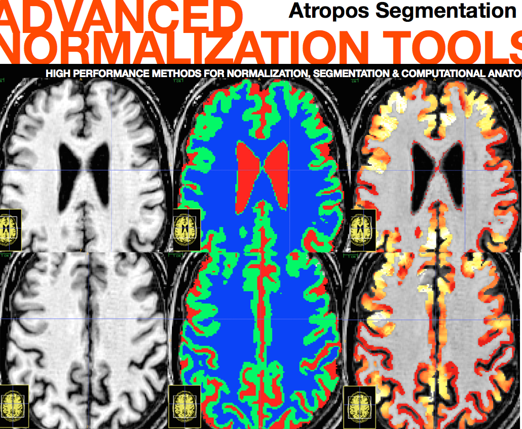
Atropos segmentation, N4 inhomogeneity correction, Eigenanatomy, SCCAN, Prior-constrained PCA, and atlas-based label fusion and MALF (powerful expert systems for segmentation)
apples and oranges …
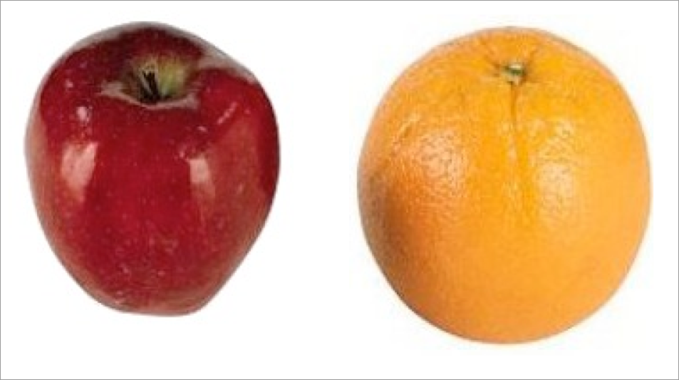
initialization
apples and oranges …
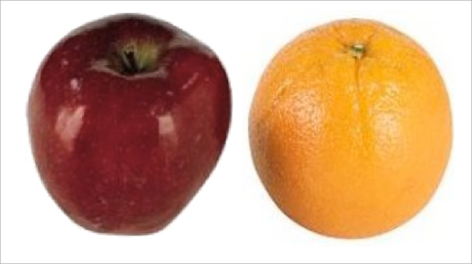
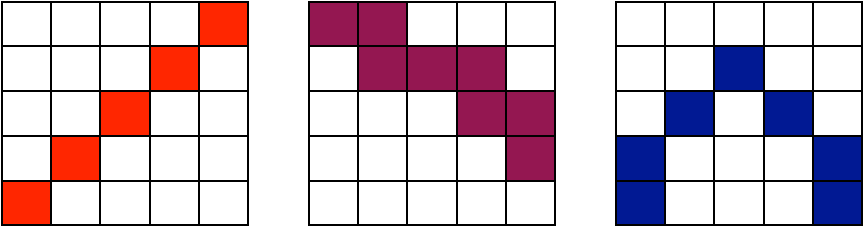
RGB affine
apples and oranges …
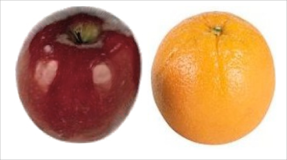
RGB deformable registration - i.e. registration on color
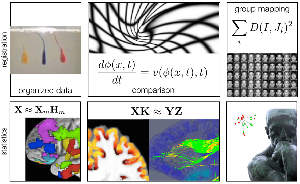
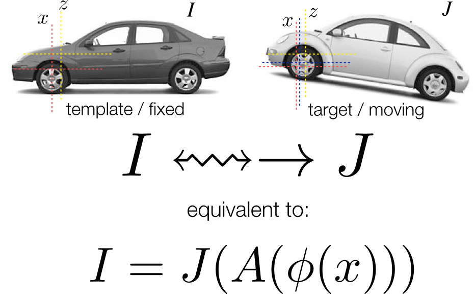
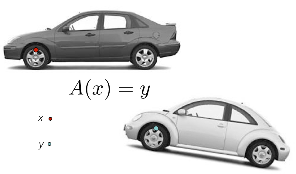
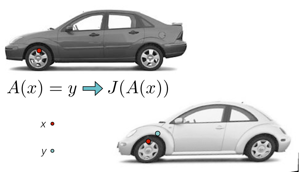
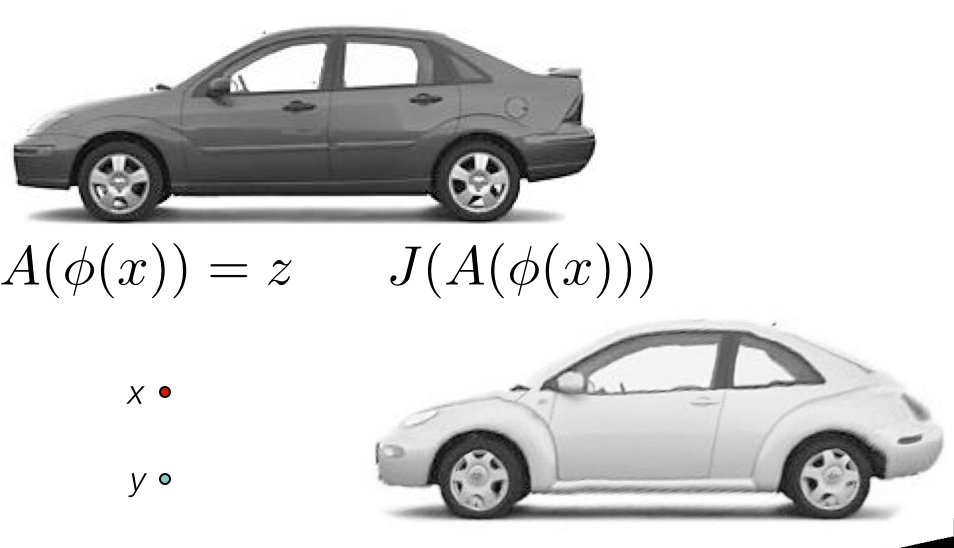
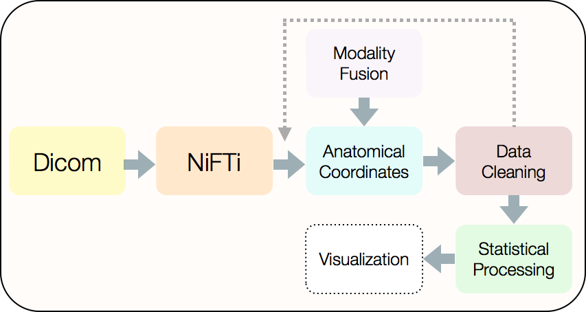
The Technical Framework
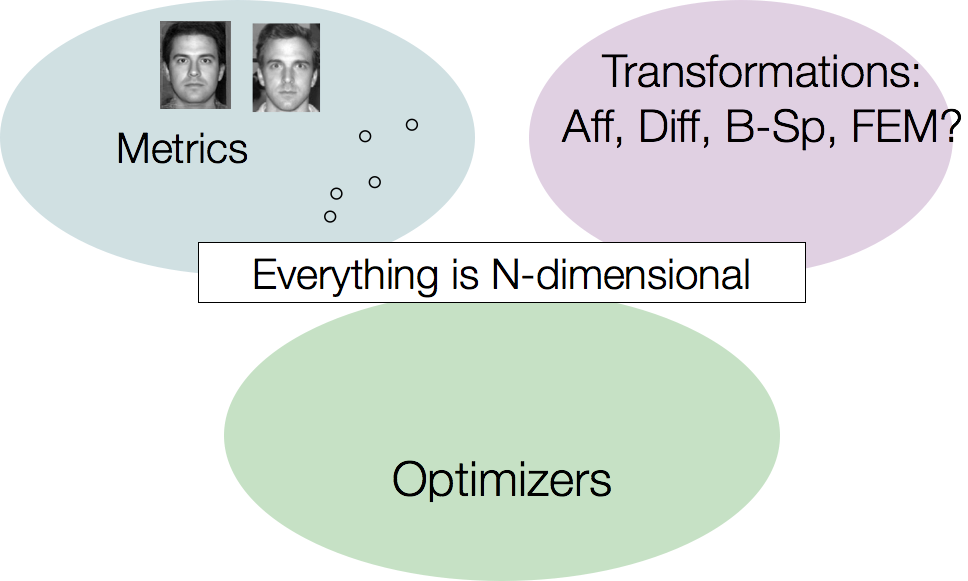
… and most of it multivariate.
ANTs Nomenclature / Standards
ANTs Nomenclature / Standards
ANTs Nomenclature / Standards
ANTs Nomenclature / Standards
The A-team of similarity metrics
\[ \| I - J \| ~~~~~~~~~~~~~~~~~~~ \frac{< I, J >}{\|I\|\|J\|} ~~~~~~~~~~~~~~~ p(I,J) log \frac{p(I,J)}{p(I)p(J)}\]
all metrics may be computed from sparse or dense samples and used with low or high-dimensional transformations
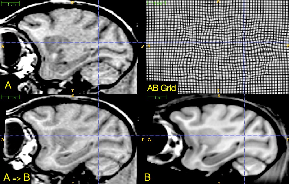
SyN for optimization symmetry
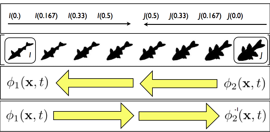 Images deform symmetrically along the shape manifold. This eliminates bias in the measurement of image differences.
Minimizing interpolations
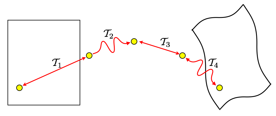
\(\mathcal{T}_{total} = \mathcal{T}_1 \circ \mathcal{T}_2 \circ \mathcal{T}_3 \circ \mathcal{T}_4\)
To avoid compounding interpolation error with the concatenation of transformations, ANTs never uses more than a single interpolation.
We ported many of these ideas into the Insight ToolKit
as part of its V4 reboot!
Sampling & feature selection: Multi-start
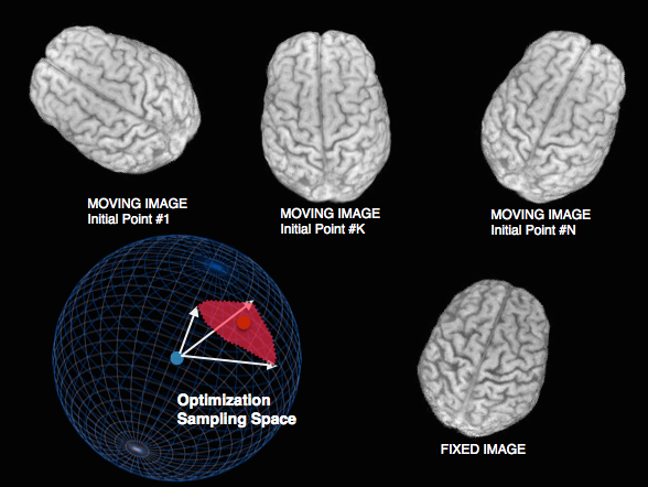
Theoretical guarantee of global optimum: improves local optimizers.
Default in antsCorticalThickness pipeline and FSL.
Sampling & feature selection: Biomedical imagery
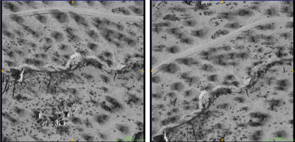
Initial configuration of data
Sampling & feature selection: Biomedical imagery
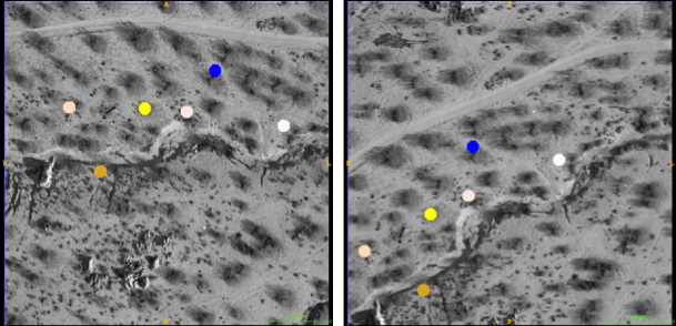
Automatic feature selection
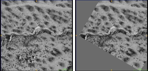
Sampling & feature selection: Biomedical imagery
Resampling allows comparison & slide alignment and
validates the feature selection
Dramatic reduction in computation time / memory requirements
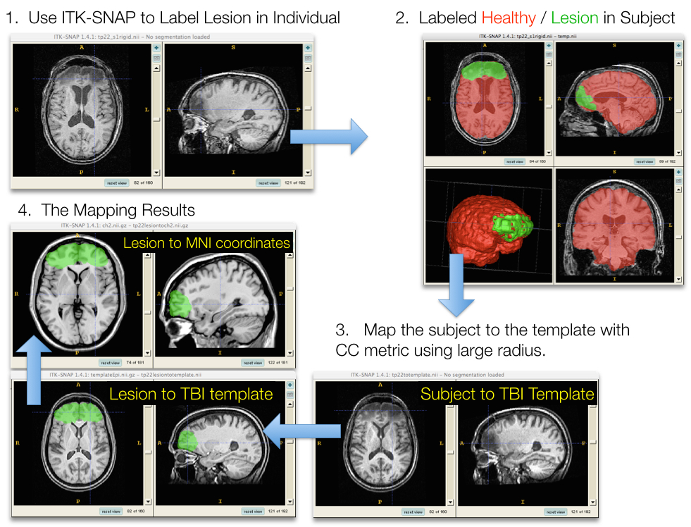
Sampling & feature selection: Lesioned brains
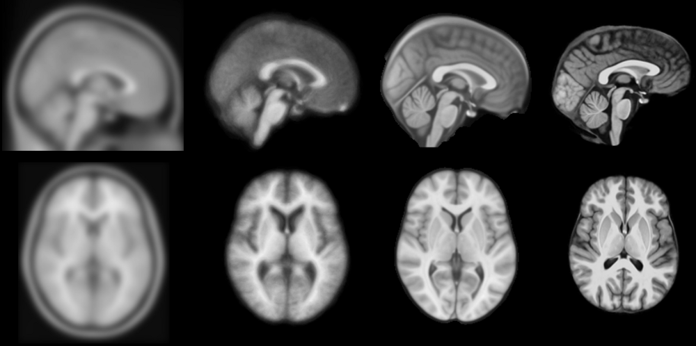
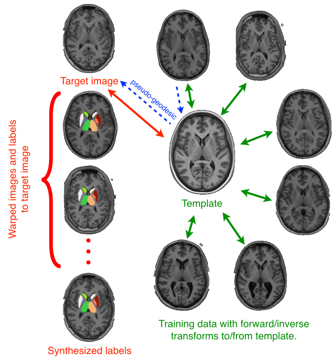
Brain templates as high-dimensional averages

SyGN - templates and averages in deformation space
 from miykael
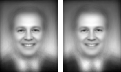
Average Republican and Democratic congressmen
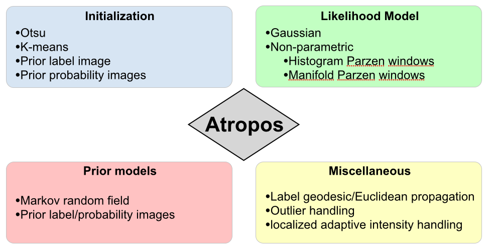
Atropos components
Babies
KellySlater \(\rightarrow\) KellyKapowski
Several years of development by SR Das, BA, NT (KK fan)
Atropos \(+\) KK Example
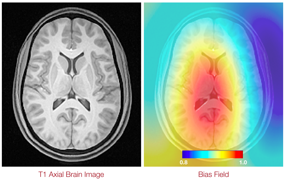
N4 Introduction
Anatomical dictionaries
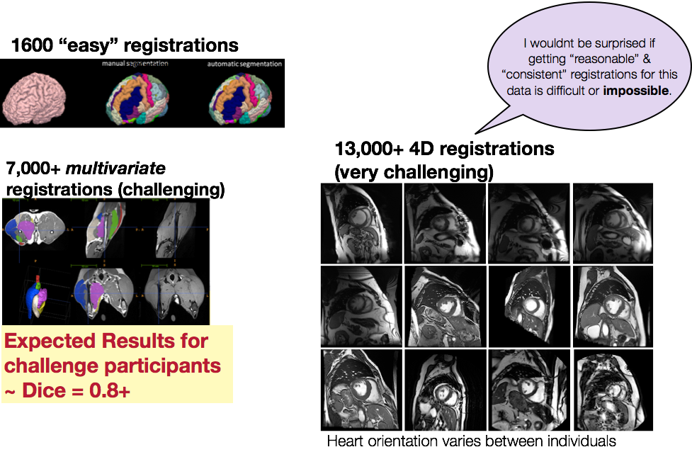
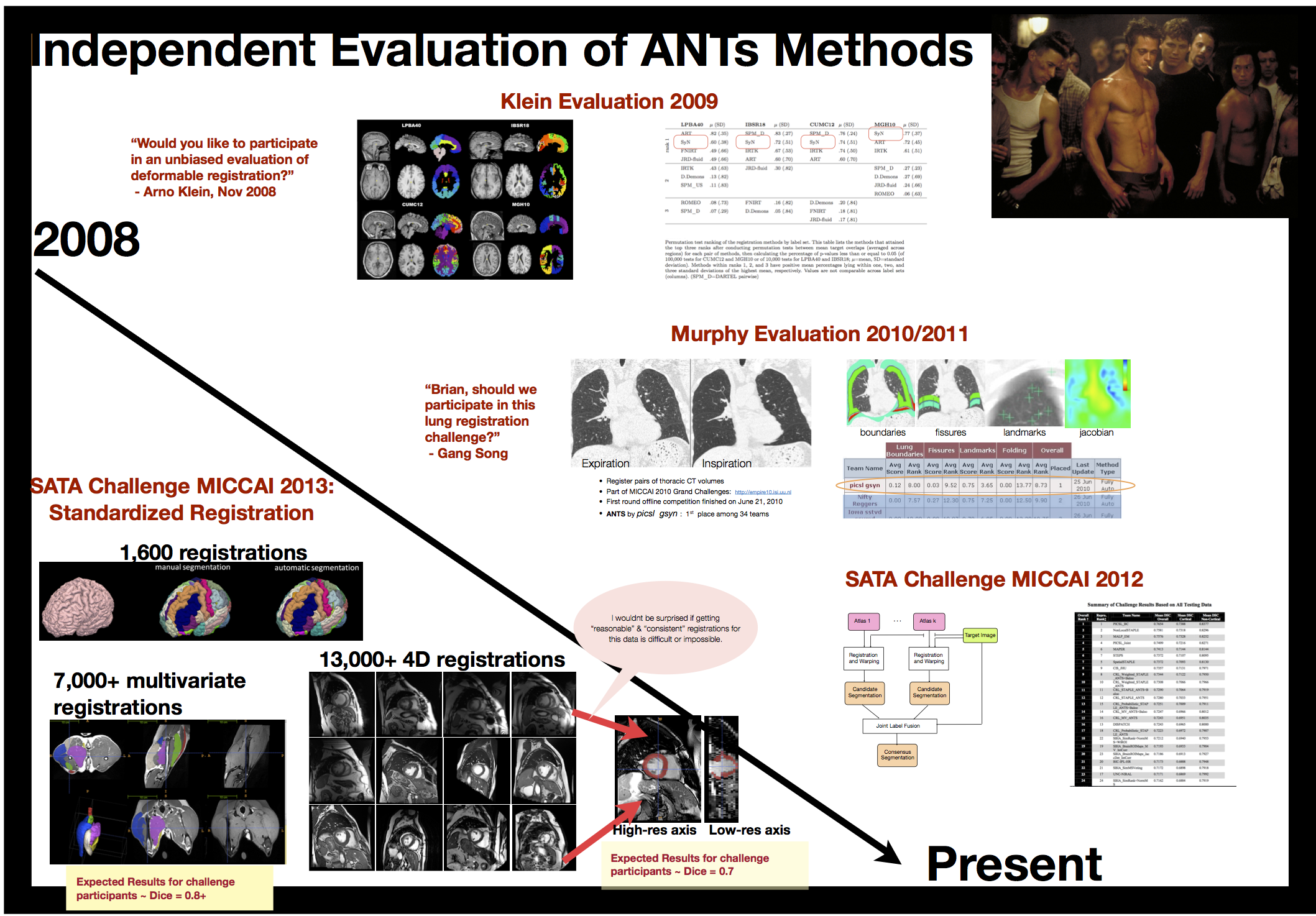
we provided the standard registration results for \(>\) 20,000 image pairs at SATA 2013
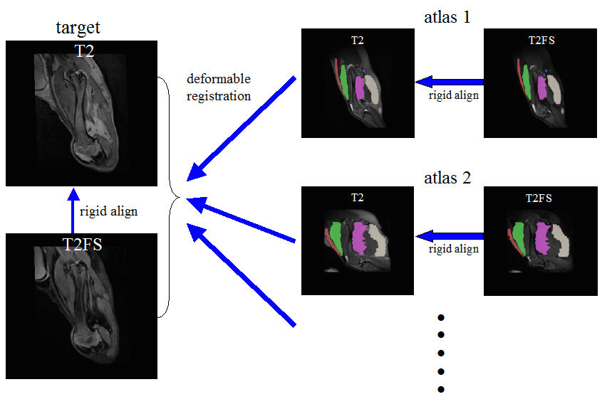
label fusion (link)
Multiple metrics improve performance
to our knowledge, ANTs is the only freely available system that can solve this problem in a fully multivariate manner.
Hongzhi Wang won the “walk in the park” award for this work …
“Big data” problem from public resources
TOT, NKI, IXI, Oasis, ADNI … several thousand images
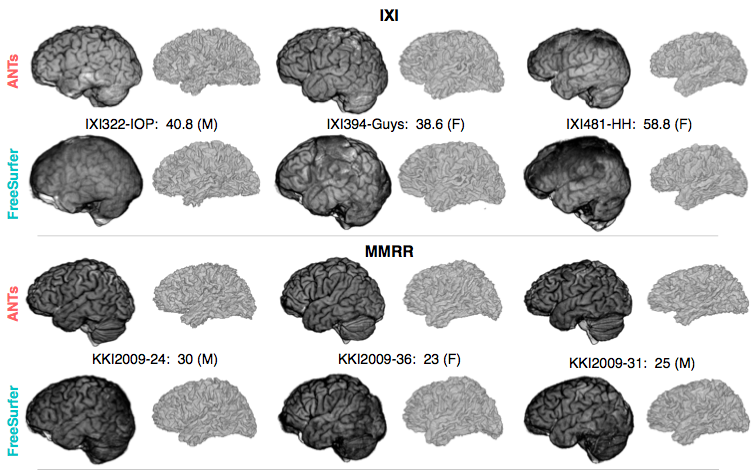
ANTS vs Freesurfer
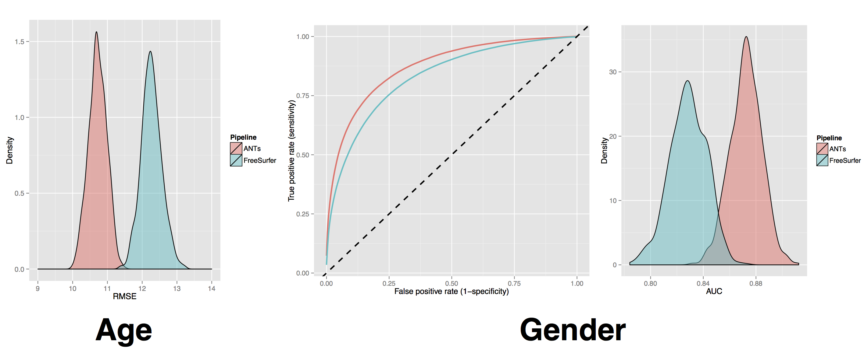
ANTs vs Freesurfer 2
ANTs MALF Labeling
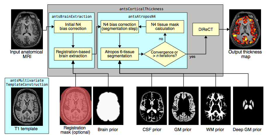
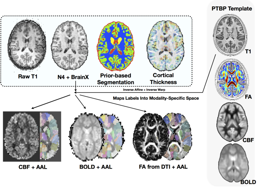
The ANTs structural brain mapping pipeline
Large-scale evaluation of ANTs* and FreeSurfer cortical thickness measurements, NeuroImage 2014.
All software components are open source and part of the Advanced Normalization Tools (ANTs) repository.
Basic components of the pipeline
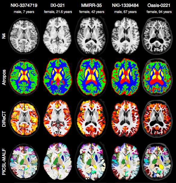
- template building (offline)
- brain extraction
- cortical thickness estimation
- cortical parcellation
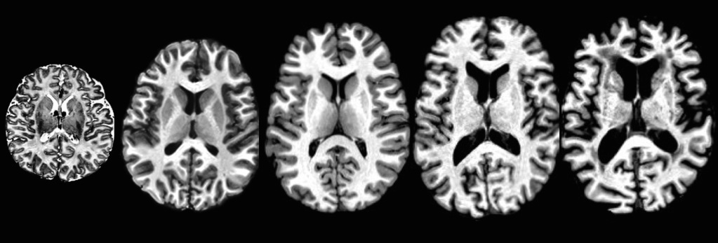
Template building
Tailor data to your specific cohort
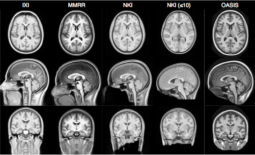
- Templates representing the average mean shape and intensity are built directly from the cohort to be analyzed, e.g. pediatric vs. middle-aged brains.
- Acquisition and anonymization (e.g. defacing) protocols are often different.
Template building (cont.)
Each template is processed to produce auxiliary images which are used for brain extraction and brain segmentation.
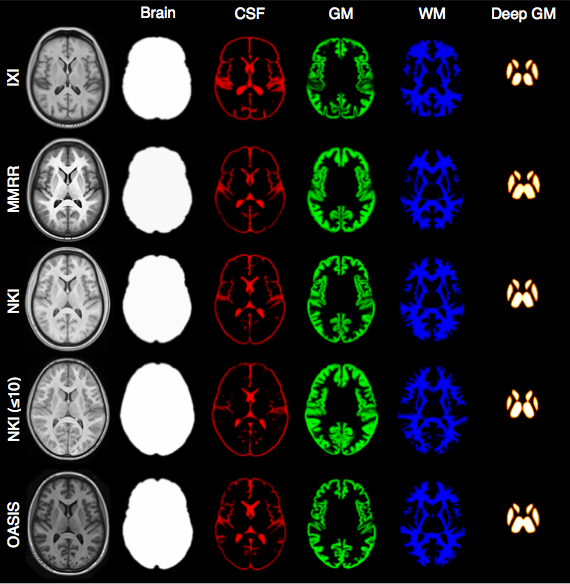
Brain extraction
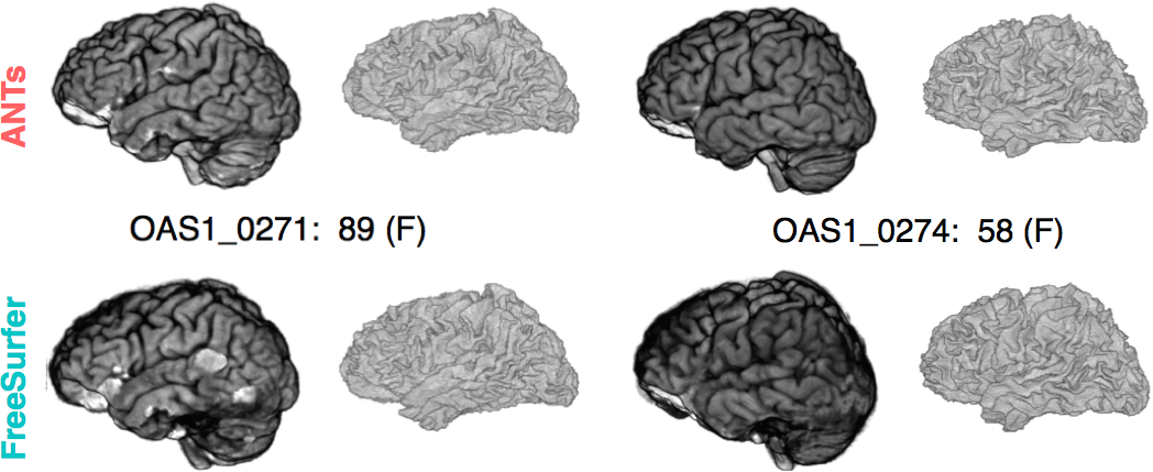
Comparison with de facto standard FreeSurfer package. Note the difference in separation of the gray matter from the surrounding CSF. (0 failures out of 1205 scans)
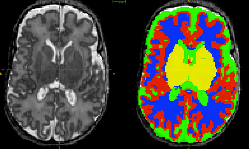
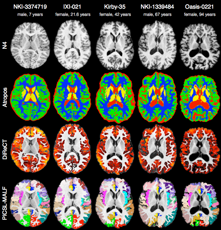
Brain segmentation
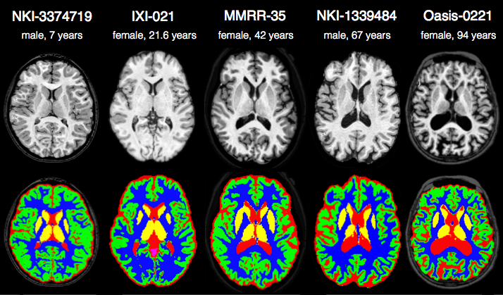
Randomly selected healthy individuals. Atropos gets good performance across ages.
Cortical thickness estimation
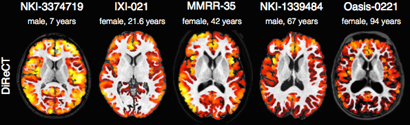
In contrast to FreeSurfer which warps coupled surface meshes to segment the gray matter, ANTs diffeomorphically registers the white matter to the combined gray/white matters while simultaneously estimating thickness.
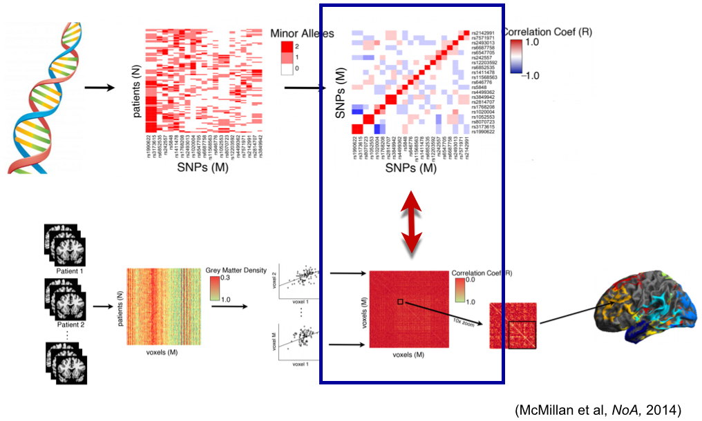
multivariate statistical fields arise from fused modalities
Many opportunities for statistical advancements
Scientific Data 2014
Agnostic statistics
Visualize the histograms of effects
whichvox<-qvals < 1.e-2
voxdf<-data.frame( volume=c( as.numeric( mat[,whichvox] ) ), DX=DX )
ggplot(voxdf, aes(volume, fill = DX)) + geom_density(alpha = 0.2)
Visualize the anatomical distribution
plotANTsImage(img,functional=list(betas),threshold=thresh,
  outname=ofn)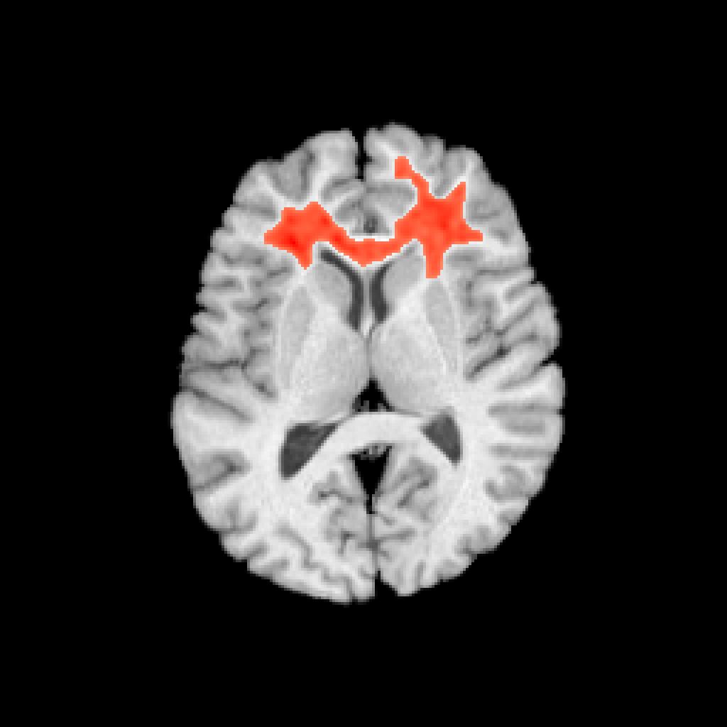
Network visualization
see ?plotBasicNetwork
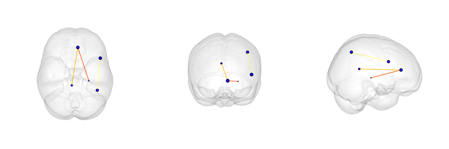
The power of ANTs \(+\) R \(\rightarrow\)
Reproducible imaging science
… used in “Sparse canonical correlation analysis relates network-level atrophy to multivariate cognitive measures in a neurodegenerative population” and several upcoming …
Tools you can use for imaging science
Core developers: B. Avants, N. Tustison, H. J. Johnson, J. T. Duda
Many contributors, including users …
Multi-platform, multi-threaded C++ stnava.github.io/ANTs
Developed in conjunction with http://www.itk.org/
R wrapping and extension stnava.github.io/ANTsR
rapid development, regular testing \(+\) many eyes \(\rightarrow\) bugs are shallow